Beranda
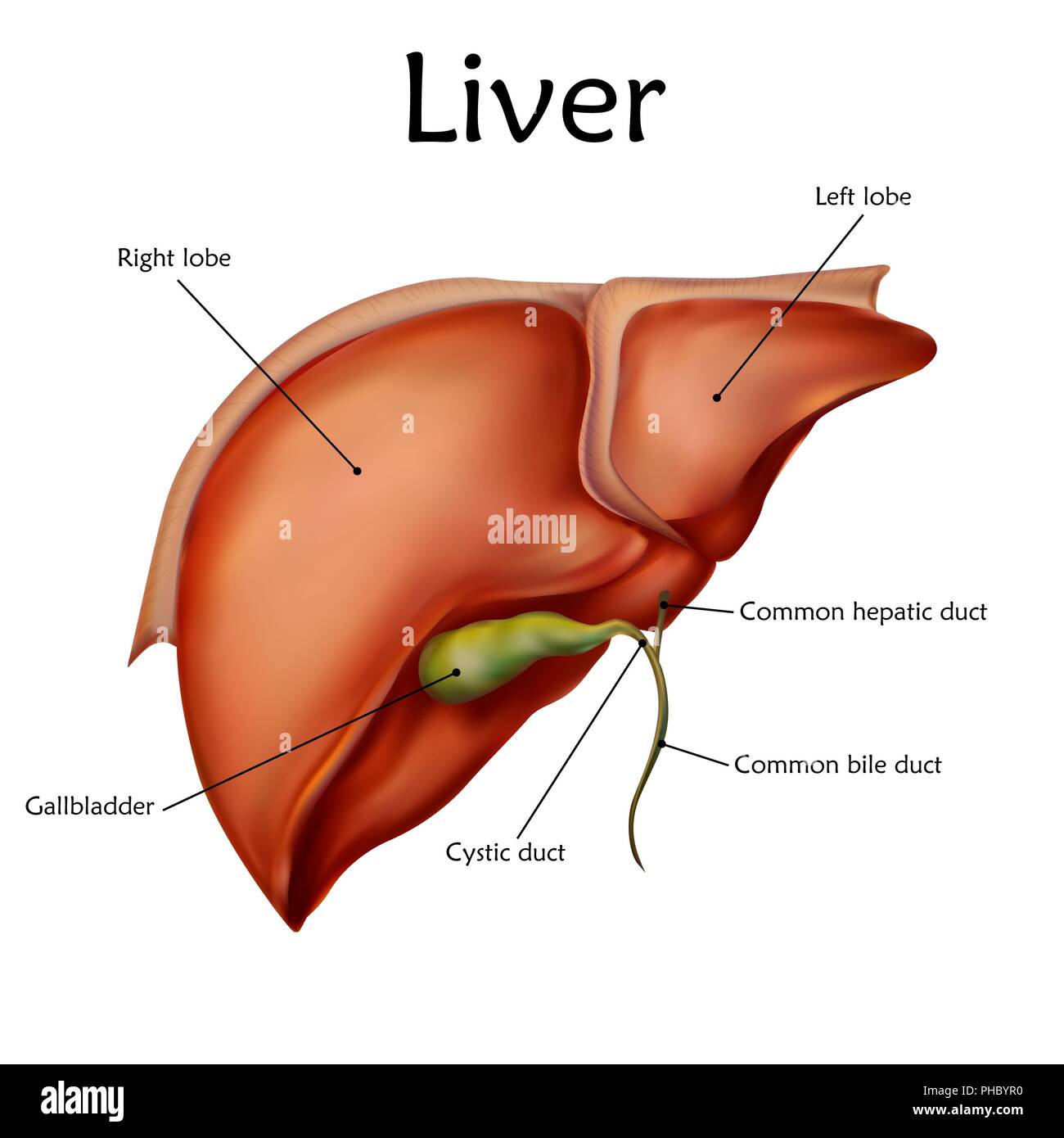
/ Liver Diagram With Labels : Liver Gallbladder And Pancreas Labeled Anatomical Illustration Stock Illustration Illustration Of Ducts Island 160724739 - Through liver diagram we can also understand the liver anatomy and liver structure clearly.
Liver Diagram With Labels : Liver Gallbladder And Pancreas Labeled Anatomical Illustration Stock Illustration Illustration Of Ducts Island 160724739 - Through liver diagram we can also understand the liver anatomy and liver structure clearly.
Insurance Gas/Electricity Loans Mortgage Attorney Lawyer Donate Conference Call Degree Credit Treatment Software Classes Recovery Trading Rehab Hosting Transfer Cord Blood Claim compensation mesothelioma mesothelioma attorney Houston car accident lawyer moreno valley can you sue a doctor for wrong diagnosis doctorate in security top online doctoral programs in business educational leadership doctoral programs online car accident doctor atlanta car accident doctor atlanta accident attorney rancho Cucamonga truck accident attorney san Antonio ONLINE BUSINESS DEGREE PROGRAMS ACCREDITED online accredited psychology degree masters degree in human resources online public administration masters degree online bitcoin merchant account bitcoin merchant services compare car insurance auto insurance troy mi seo explanation digital marketing degree floridaseo company fitness showrooms stamfordct how to work more efficiently seowordpress tips meaning of seo what is an seo what does an seo do what seo stands for best seotips google seo advice seo steps, The secure cloud-based platform for smart service delivery. Safelink is used by legal, professional and financial services to protect sensitive information, accelerate business processes and increase productivity. Use Safelink to collaborate securely with clients, colleagues and external parties. Safelink has a menu of workspace types with advanced features for dispute resolution, running deals and customised client portal creation. All data is encrypted (at rest and in transit and you retain your own encryption keys. Our titan security framework ensures your data is secure and you even have the option to choose your own data location from Channel Islands, London (UK), Dublin (EU), Australia.
Liver Diagram With Labels : Liver Gallbladder And Pancreas Labeled Anatomical Illustration Stock Illustration Illustration Of Ducts Island 160724739 - Through liver diagram we can also understand the liver anatomy and liver structure clearly.. Drag the labels onto the diagram to identify the anatomical features of the liver. Several diagrams of liver structure removed for copyright reasons. Bio 234 lab unit ii a detailed description of the liver: Anatomically the liver consists of four lobes. Digestive systems picture and label 12 photos of the digestive systems picture and label digestive system picture and labels, digestive system picture to label, digestive system picture with label, digestive system picture without label, human digestive system picture with label, inner body, digestive system.
Although segment iv is part of the left hemiliver, it is situated more to the right. Human liver diagram with labels for exam point of perspective. The label titled pharynx was pointing to the uvula so this was corrected by correctly labeling pointers to both the uvula and the pharynx. The cell membrane is the outer coating of the cell and contains the cytoplasm substances within it and the organelle. The hepatic artery and the portal vein.
What Does The Liver Produce Labeled Diagram Royalty Free Cliparts Vectors And Stock Illustration Image 32265270 from previews.123rf.com And also you can download liver images from here. Uruj zehra mbbs, mphil, phd last reviewed: The portal vein and the. Liver, gallbladder and pancreas, labeled, anatomical illustration. Bio 234 lab unit ii a detailed description of the liver: Anatomically the liver consists of four lobes. The nomenclature of some of the ligaments is based on the structures that they connect, so it's quite easy to remember them. Describe the function of the different parts of the digestive system.
Drag the labels onto the diagram to identify the anatomical features of the liver.
The hepatic artery and the portal vein. Most of the liver's mass is located on the right side of the body where it descends inferiorly toward the right kidney. There are 2 distinct sources that supply blood to the. February 23, 2021 reading time: Animal cell diagram given the label on diagram identify the cell part. Illustration digestive system vector illustration. Bio 234 lab unit ii a detailed description of the liver: This update is an attempt to correct that. Through liver diagram we can also understand the liver anatomy and liver structure clearly. One abnormal characteristic is the liver's regenerative abilities. Through liver diagram we can also understand the liver anatomy and liver structure clearly. Several diagrams of liver structure removed for copyright reasons. Couinaud divided the liver into a functional left and right liver by a main portal scissurae containing the middle hepatic vein.
Cells that are rapidly undergoing mitosis constantly repair and renew the lining of the pharynx and the esophagus, which is particularly vulnerable to abrasion associated with swallowing. The label titled pharynx was pointing to the uvula so this was corrected by correctly labeling pointers to both the uvula and the pharynx. This update is an attempt to correct that. Illustration boy and liver on diagram. The portal vein and the.
Human Liver With Labels Illustration Stock Photo Alamy from c8.alamy.com This is an online quiz called liver, pancreas, gallbladder anatomy there is a printable worksheet available for download here so you can take the quiz with pen and paper. Describe the function of the different parts of the digestive system. Reset help bile duct right lobe inferior vena cava hepatic artery proper caudate lobe left lobe gallbladder hepatic portal vein quadrate lobe hepatic vein coronary ligament coronary ligament left lobe porta hepatis right lobe falciform ligament round ligament gallbladder the anterior surface of the liver the. The hepatic artery and the portal vein. Figures 2 and 3 in harada, t., et al. Although segment iv is part of the left hemiliver, it is situated more to the right. One abnormal characteristic is the liver's regenerative abilities. Mouth, pancreas, stomach, gallbladder, small intestine, oesophagus, large intestine, anus, liver.
Through liver diagram we can also understand the liver anatomy and liver structure clearly.
Most of the liver's mass is located on the right side of the body where it descends inferiorly toward the right kidney. Vector illustration of liver cancer diagram in detail. This is an online quiz called liver, pancreas, gallbladder anatomy there is a printable worksheet available for download here so you can take the quiz with pen and paper. Mouth, pancreas, stomach, gallbladder, small intestine, oesophagus, large intestine, anus, liver. Couinaud divided the liver into a functional left and right liver by a main portal scissurae containing the middle hepatic vein. Drag the labels onto the diagram to identify the anatomical features of the liver. Adrian rad bsc (hons) • reviewer: Drag and drop the pins to their correct place on the image. Human liver diagram with labels for exam point of perspective. The portal vein and the. This is known as cantlie's line. Animal cell diagram given the label on diagram identify the cell part. The right border of the liver is formed by segment v and viii.
There are 2 distinct sources that supply blood to the. Describe the function of the different parts of the digestive system. This is known as cantlie's line. Drag and drop the pins to their correct place on the image. Liver, gallbladder and pancreas with hepatic ducts, cystic duct, bile duct, duodenum, pancreas.
Https Encrypted Tbn0 Gstatic Com Images Q Tbn And9gcqlyq1gtyvcognxk3763qkgzzysm Bfnflhbf5pzpupdb3bboxi Usqp Cau from However, it can be felt ascending and descending if you. Describe the function of the different parts of the digestive system. Drag the labels onto the diagram to identify the anatomical features of the liver. This is the well labelled diagram of liver fluke. Identify the main parts and describe their function. The hepatic artery and the portal vein. There are 2 distinct sources that supply blood to the. Illustration boy and liver on diagram.
Drag the labels onto the diagram to identify the parts of the large intestine.
In both animals and plants cells generally become specialized to perform certain functions. The liver parenchyma consists of a complex network of epithelial cells, supported by connective tissue, and perfused by a rich blood supply from the hepatic portal vein and hepatic artery. How to draw a liver fluke in exam is the topic. This is the well labelled diagram of liver fluke. The right border of the liver is formed by segment v and viii. This update is an attempt to correct that. Liver, gallbladder and pancreas with hepatic ducts, cystic duct, bile duct, duodenum, pancreas. The cell membrane is the outer coating of the cell and contains the cytoplasm substances within it and the organelle. Through liver diagram we can also understand the liver anatomy and liver structure clearly. 595 × 842 (205 kb) dbrouse~commonswiki: Related posts of 3d diagram of human liver digestive systems picture and label. The label titled pharynx was pointing to the uvula so this was corrected by correctly labeling pointers to both the uvula and the pharynx. Uruj zehra mbbs, mphil, phd last reviewed:
Related posts of 3d diagram of human liver digestive systems picture and label liver diagram. The portal vein and the.